
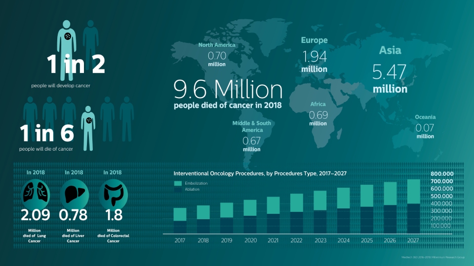
A global trend in oncology care
Many minimally-invasive, imaging-guided procedures are replacing more traditional open surgical techniques for treating solid tumors in a variety of organs – most often in the liver (primary and metastatic tumors), lung and kidney [1].
Originally considered the therapy of last resort, interventional oncology has been a fast growing field for the last decade and is emerging as a recognized interventional radiology sub-specialty.
Onco suite
Tumor embolization
Adoption of chemo/radioembolization techniques such as TACE and SIRT drives the need for standardization and efficiency. Case after case, you must reliably and consistently locate the tumor(s), identify all feeder vessels, and plan/execute the appropriate interventional approach.
Tumor ablation
Percutaneous ablation procedures are increasingly performed by interventional radiologists, and adoption may accelerate the shift away from open surgery. The key is gaining a clear understanding of the tumor size, needle ablation area, and optimal path to the target.
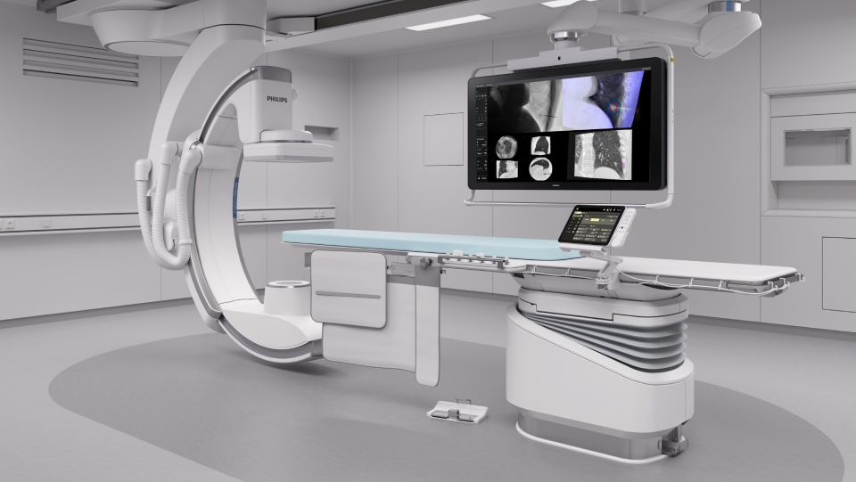
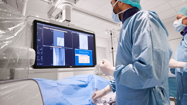
Philips Onco suite for the confident path to treatment
Interventional oncology has evolved from a niche specialty to a well-established treatment alternative for various types of cancers. Strong clinical evidence supports procedures such as transcatheter embolization for primary liver cancer and metastases, and focal tumor ablation therapy.
Onco suite is a combination of the Azurion platform, interventional solutions, workflow options, education, and services.
Visualize full tumor enhancement and feeding arteries through dual phase CBCT acquisition, part of SmartCT soft tissue.
Overlay your pre-procedural PET/MR or CT images for optimal needle path planning with XperGuide.
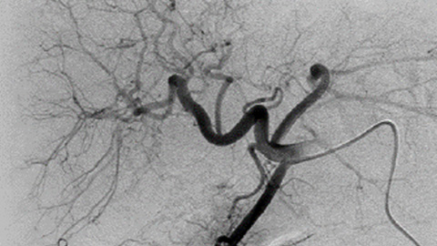
Depict all tumor feeders with EmboGuide’s automatic detection and navigate easily to your targeted arteries with live guidance.
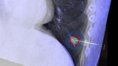
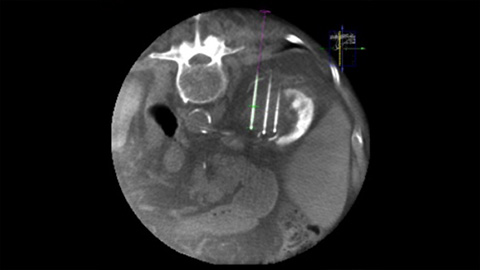
Optimize your needle position and ablation coverage with live guidance and virtual ablation zone visualization.
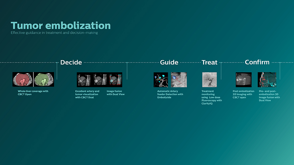
Tumor embolization
Adoption of chemo/radioembolization techniques such as TACE and SIRT drives the need for standardization and efficiency. Case after case, you must reliably and consistently locate the tumor(s), identify all feeder vessels, and plan/execute the appropriate interventional approach. Onco suite provides tools to help you:
Easy guided CBCT acquisition with SmartCT*
SmartCT is designed to make 3D imaging accessible to all clinical users** regardless of their level of experience. SmartCT offers step by step guidance throughout the whole 3D image acquisition leaving you no room for guess work and empowering you to deliver superior care to your patients.
Total table side control with SmartCT*
Easily control advanced 3D acquisition, visualization and measurements at table side to improve lab flexibility and efficiency. Segmentation tasks are semi-automated and made available on the touch screen to speed up your 3D image analysis. SmartCT gives you total control of 3D imaging at table side while remaining in the sterile field which can potentially help you save time during your procedures.
Whole liver coverage with CBCT Open
By opening the arc to the left of the patient, CBCT allows off center positioning of the patient table and therefore better centering of the FOV. It significantly increases image coverage to help visualize tumors on the periphery of the organ [2].
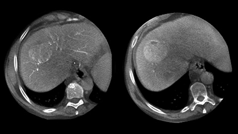
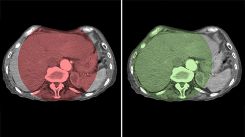
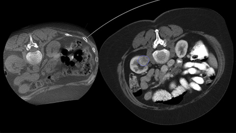
Optimal artery and tumor visualization with CBCT Dual
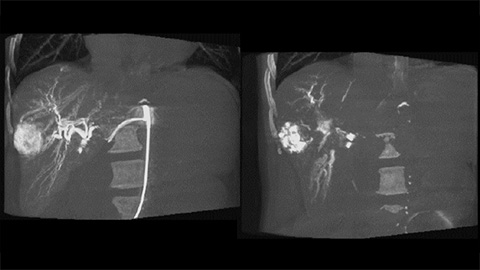
CBCT enables 3D acquisition of an arterial phase to visualize vascular structures and a post-arterial (delayed phase) to optimally visualize accumulation of contrast medium, in a single automatic step [3].
Image fusion with Dual View
Dual View allows simultaneous visualization of two CBCT datasets (pre and post). Both arterial and delayed phase can be displayed next to each other or in a single fused overlay view.
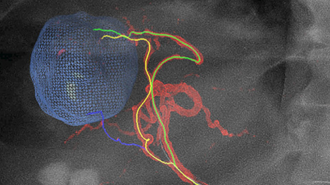
Automatic Artery feeder Detection with EmboGuide
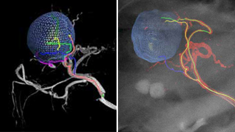
EmboGuide supports you in maximizing the efficacy of your TACE procedures as it potentially enhances your sensitivity, reduces false positives and maximizes inter-reader agreement [4]. It provides efficient, workflow-based live 3D guidance with automatic feeder detection [5].
Treatment monitoring using low dose fluoroscopy with ClarityIQ
ClarityIQ produces tuned, high definition low dose images with superb vascular detail to monitor the embolization [6].
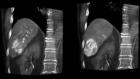
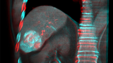
Post embolization 3D imaging with CBCT
A post embolization 3D acquisition allows you to visualize the targeted deposition of embolic material, such as Liopidol or radiopaque beads, in the tumor [7].
Pre-and Post-embolization 3D image fusion with Dual View
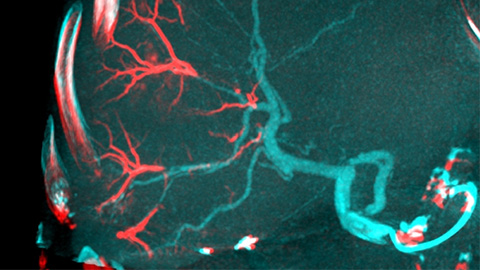
Dual View allows simultaneous visualization of pre-embolization arterial phase 3D image and the post embolization image to assess treatment endpoint and predict outcome.
Ablation
Percutaneous ablation (radiofrequency, microwave, and cryoablation) is a well-established minimally invasive treatment of kidney, liver, lung and bone tumors. The key is gaining a clear understanding of the tumor size, needle ablation area, and optimal path to the target. Onco suite provides tools to help you:
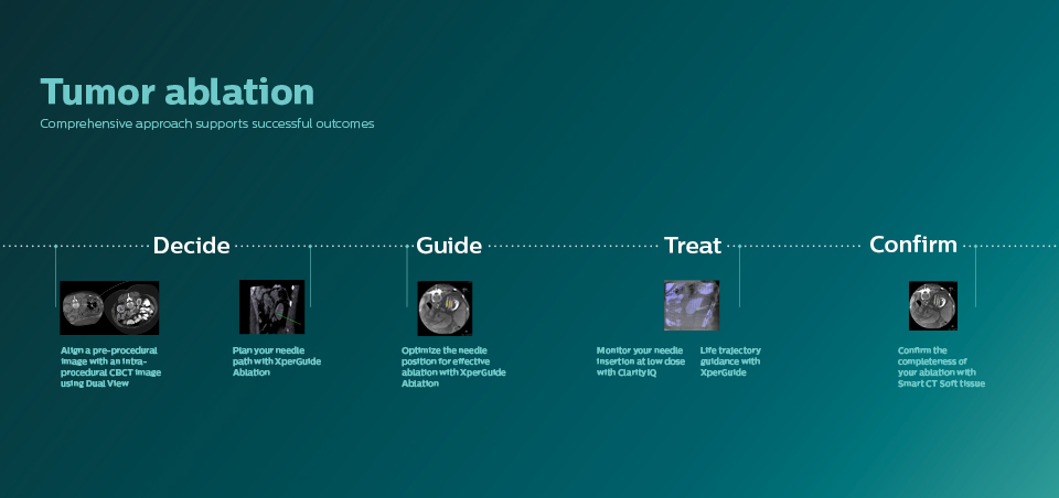
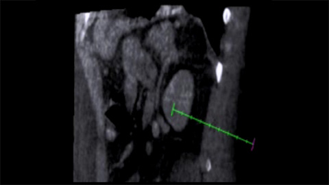
Align a pre-procedural image with a new CBCT 3D image using Dual View
Dual View allows the overlay of a pre-procedure 3D image (CT/MR/PET-CT) on an intra-procedure 3D CBCT to better visualization the lesions and access critical input for needle planning [8].
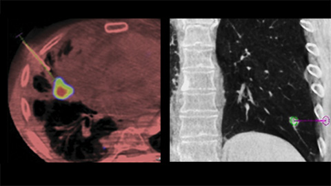
Plan your needle path with XperGuide Ablation
XperGuide Ablation provides comprehensive assistance for treatment planning and live needle guidance. It offers unique Parallax Correction to plan needle trajectories for off-center lesions [9].
Optimize the needle position for effective ablation with XperGuide Ablation
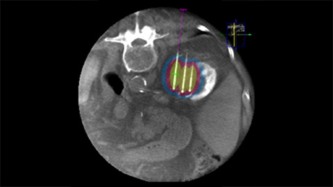
XperGuide Ablation displays the virtual needle path to assist in multiple needle planning. It shows needle characteristics such as ablation zone/isotherm to confirm complete tumor coverage prior to ablation.
Monitor your needle insertion at low dose with ClarityIQ
Clarity IQ produces finely tuned high-definition fluoroscopic images with superb detail to assist with needle progression to target [6].
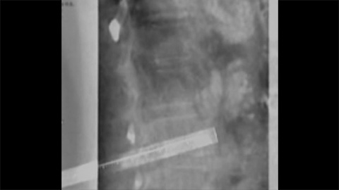
Live trajectory guidance with XperGuide
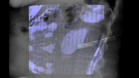
XperGuide provides highly accurate live image guidance of each needle to a targeted position by overlaying pre-planned trajectories with fluoroscopic imaging [10].
Confirm the completeness of your ablation with CBCT
With CBCT acquire a post-ablation CBCT to demonstrate the extent of tumor coverage and confirm completeness of your treatment.
Optimize lab performance and dose management
Work with confidence.
Azurion Onco suite offers a number of workflow innovations designed to help oncology teams work efficiently and consistently, while maintaining a single-minded focus on the patient and keeping radiation dose low during interventions.
-
-
Zero Dose Positioning
This features helps you manage dose by allowing you to pan the table, change table height or move the X-ray system on Last Image Hold to determine the new center position. This helps you prepare your next run without using fluoroscopy.You are about to visit a Philips global content page
Continue -
20" detector
High-resolution imaging over a large field of view with full projection flexibility.You are about to visit a Philips global content page
Continue -
Touch screen module Pro
Allows table side control of images and applications with tablet ease to save time and unnecessary walking in and out of the sterile area.
You are about to visit a Philips global content page
Continue -
FlexVision Pro
Gives you full control of all system inputs including CX50 ultrasound for your soft tissue biopsies at tableside to save time and unnecessary walking in and out of the sterile area.
You are about to visit a Philips global content page
Continue -
ClarityIQ technology
ClarityIQ X-ray imaging technology provides superb image quality at significantly lower dose across clinical areas, patients, and Operators. One study showed that ClarityIQ reduces patient dose by 83% in iliac DSA procedures, while maintaining equivalent image quality, compared to a system without ClarityIQ.
You are about to visit a Philips global content page
Continue
17% reduction of procedure time with Philips Azurion at St. Antonius Hospital11
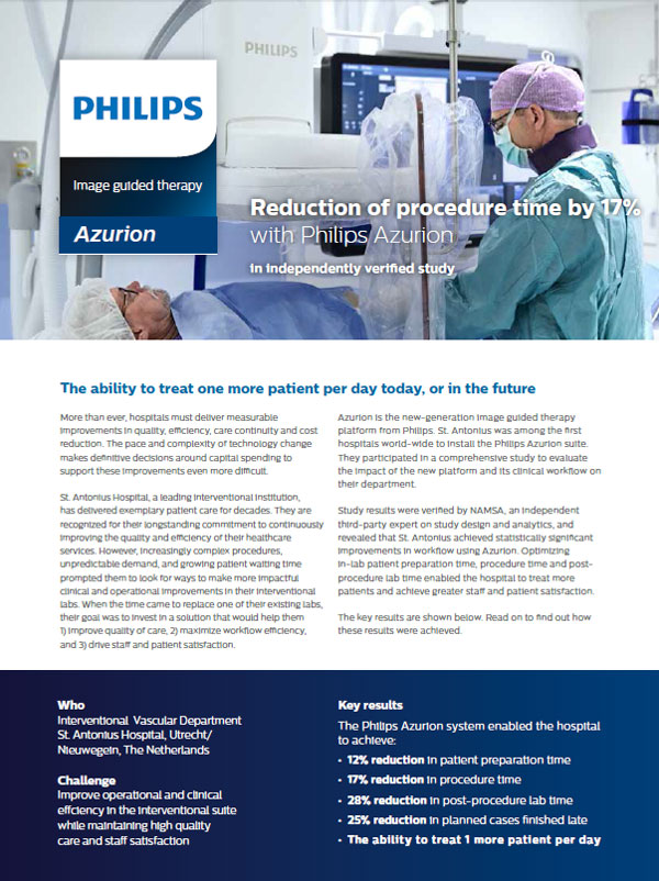
The ability to treat one more patient per day, or in the future
This is just one of the many improvements in lab performance achieved by the Interventional Vascular Department at St. Antonius Hospital after installing the Azurion system. The first Azurion lab performance study achieved impressive results which have been verified by an independent third party.
ClarityIQ technology – clinically proven
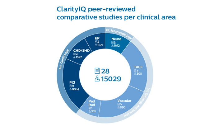
ClarityIQ X-ray imaging technology provides superb image quality at significantly lower dose across clinical areas, patients, and Operators12.
FlexVision Pro monitor
With FlexVision Pro, you can intuitively control all available applications at tableside via seamless mouse over to streamline your workflow.
Touch screen module Pro
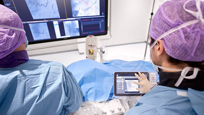
With touch screen module Pro, you can easily control system settings and applications from Philips and other vendors, plus you can control the X-ray image for easy navigation – all at tableside. It works just like a tablet to help you work quickly and easily.
FlexSpot – flexible workspot
Team members can control all connected applications in the control room with one keyboard and mouse. Screen layout can be customized by user preferences and items can be re-sized, dragged and dropped on the fly. FlexSpot reduces clutter and simplifies workflow.
Zero Dose Positioning to manage dose
This features helps you manage dose by allowing you to pan the table, change table height or move the X-ray system on Last Image Hold to determine the new center position. This helps you prepare your next run without using fluoroscopy.
ProcedureCards standardize system setup
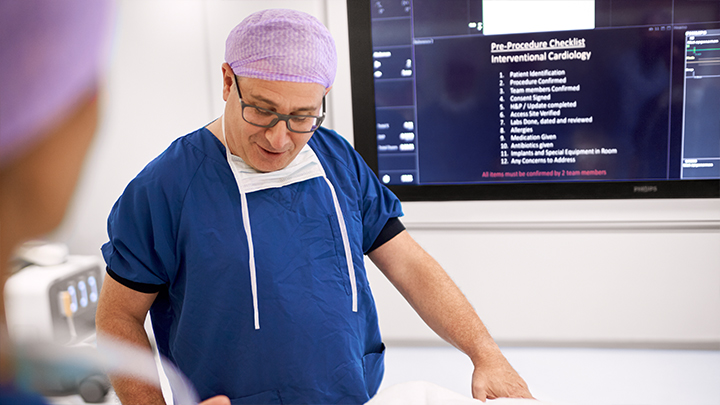
ProcedureCards help you streamline and standardize system set-up and reduce preparation errors. Select the TACE ProcedureCard and the system is set-up the way you want. Hospital specific protocols and/or checklists can be added to ProcedureCards and displayed on monitors to support consistent workflow.
*SmartCT is 510(k) pending in the U.S.A. Not available for sales in the U.S.A ** As described in the SmartCT Instructions for Use 1. Medtech 360 2016-2019, Millennium Research Group 2. Schernthaner RE et al, Feasibility of a Modified Cone-Beam CT Rotation Trajectory to Improve Liver Periphery Visualization during Transarterial Chemoembolization, Radiology. 2015; 277(3):833–4. 3 - Loffroy R, et al. Comparing the detectability of hepatocellular carcinoma by C-arm dual-phase cone-beam computed tomography during hepatic arteriography with conventional contrast-enhanced magnetic resonance imaging. Cardiovasc Intervent Radiol. 2012;35(1):97-104. 4. Chiaradia et al, J J,Sensitivity and Reproducibility of AFD Software for HCC, Vasc Interv Radiol 2018;29:425-431. 5. Miyayama S, et al. Identification of small hepatocellular carcinoma and tumor-feeding branches with cone-beam CT guidance technology during transcatheter arterial chemoembolization. J Vasc Interv Radiol. 2013; 24(4):501-8. 6. Trunz L. et al, Investigation of Radiation Dose Estimates and Image Quality Between Commercially Available Interventional Fluoroscopy Systems for Fluoroscopically Guided Interventional Procedures, Academic Radiology, in press, 2020 7. Levi E.B. et al, First Human Experience with Directly Image-able Iodinated Embolization Microbeads, Cardiovascular and interventional radiology, vol 39, issue 8, 1177-1186, 2016 8. Floridi C et al, Percutaneous needle biopsy of mediastinal masses under C-arm conebeam CT guidance: diagnostic performance and safety, Medical Onocology, 2017; 34(4): 1-7 9. Abi-jaoudeh et al , Cone Beam vs Conventional CT Navigation for Image-Guided Biopsy, J VascIntervRadio; 2016; 27: 1342–1349. 10. Percutaneous transthoracic needle biopsy of small (1 cm) lung nodules under C-arm cone-beam CT virtual navigation guidance; Ji Yung Choo Eur Radiol; 2013; 23:712–719. 11. Philips whitepaper 4522 991 30501; Reduction of procedure time by 17% with Philips Azurion in independent verified study. Results are specific to the institution where they were obtained and may not reflect the results achievable at other institutions. 12. In 18 individual comparative studies, Philips ClarityIQ was associated with reductions in patient radiation exposure. For the full list of clinical peer-reviewed papers go to https://www.philips.com.au/healthcare/resources/landing/alluraclarity-clinically-proven
The results of the application of dose reduction techniques will vary depending on the clinical task, patient size, anatomical location and clinical practice. The interventional radiologist assisted by a physicist as necessary has to determine the appropriate settings for each specific clinical task. Results based on DSA dose area product per frame from a single center prospective randomized study on 48 patients. DSA runs for Allura Xper with ClarityIQ and Allura Xper without ClarityIQ were randomly acquired on the same patient under same condition of geometry, field of view and injection protocol. Image quality was based on subjective assessment (side-by-side, equal or superior than the other, blinded review by 5 independent radiologists).